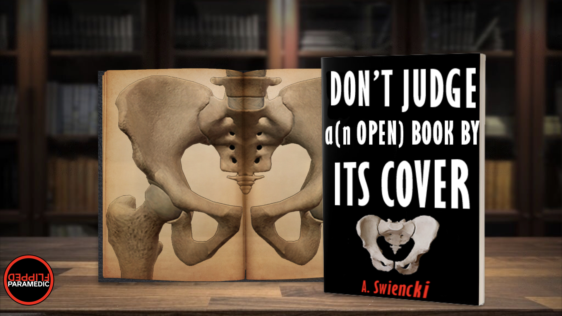
As medical providers we’re pretty good at a lot of things… identifying pelvic instability in trauma is generally not one of those things. Turns out, nobody without x-ray vision is very good at it and many fractures go unidentified. In-hospital providers depend on imaging to confidently identify or rule out pelvic fractures, but as pre-hospital providers we don’t have that luxury.
[/et_pb_text][/et_pb_column][/et_pb_row][et_pb_row _builder_version=”4.19.4″ _module_preset=”default” global_colors_info=”{}”][et_pb_column type=”4_4″ _builder_version=”4.19.4″ _module_preset=”default” global_colors_info=”{}”][et_pb_text _builder_version=”4.19.4″ _module_preset=”default” text_text_color=”#000000″ text_font_size=”20px” global_colors_info=”{}”]So, why do we care?
Pelvic fractures represent 3% or so of all skeletal injuries. Open pelvic fractures account for only 2-4% of that 3% but they’re associated with a mortality rate of up to 45%! Granted, the mechanism to sustain such an injury is pretty substantial – so there are likely concomitant injuries that contribute to that mortality rate. But in trauma patients overall, pelvic fractures are associated with an increased risk of death. Interestingly, the life threatening bleeding that occurs with these fractures is mostly venous – arterial culprits only account for about 10% of pelvic bleeding.
The average adult pelvis can hold approximately 1.5 liters of volume. But get this – the potential space created with pelvic ring disruption could accommodate the entire blood volume of the patient WITHOUT providing tamponade! That’s like 5 liters of blood, folks!
How do we properly assess for an open pelvic fracture (open book)?
Let’s start with what we don’t (or, shouldn’t) do. Those of us who’ve been in the game awhile probably remember being taught to “rock” or “spring” the pelvis. (If you’re not familiar, I’m surely not going to instill bad habits here!). These maneuvers can be dangerous, but they also provide poor sensitivity and specificity – so, risk >>> reward.
Instead, when the pelvis of a trauma patient is assessed it should be gentle; pressure should be directed posterior and medial, and it should be done ONCE. The last thing we want to do is further displace bone fragments or shear vessels or exacerbate injuries. The goal is to examine in a way where we would feel the fracture closing between our hands, we don’t want to further displace the injury (i.e. “open the book” even more).
But wait… I thought we stink at identifying pelvic fractures?
No offense, but the numbers aren’t exactly in our favor. In one study of 7201 patients with partially stable and unstable fractures, the fractures were missed 40.5% and 32.3% of the time respectively. A recent review and meta-analysis of studies (where physicians examined the patients in hospitals) found that imaging should be performed regardless of physical exam or level of consciousness. Obviously we don’t have the ability to scan in the prehospital environment – so we should take similar precautions and assume pelvic injury when the possibility is present.
So, who needs pelvic stabilization?
If the mechanism exists and your patient can’t tell you it doesn’t hurt when you perform your (gentle) pelvic exam, bind them. You don’t need to wait for hemodynamic instability, but that should be another check in the “do it” column. The potential risks of missing an occult pelvic injury outweigh the risks of pelvic binding.
Who doesn’t need it?
In our higher GCS patients (generally >13), physical exams are highly specific for fractures.
Notwithstanding other factors, if you can trust your patient, you don’t have to bind these guys if they don’t have pain.
Also, patients with isolated falls from standing height with hip or pelvic pain. These falls usually result in hip fractures (femoral neck or trochanter), not ring disruption. Applying the binder may cause further hip fracture and pain. If you’re not convinced your patient needs the binder, but also not convinced they don’t – you can place the binder on the patient and just not tighten. If your patient starts experiencing hemodynamic compromise, have a low threshold to tighten it up
[/et_pb_text][/et_pb_column][/et_pb_row][et_pb_row column_structure=”1_2,1_2″ _builder_version=”4.19.4″ _module_preset=”default” global_colors_info=”{}”][et_pb_column type=”1_2″ _builder_version=”4.19.4″ _module_preset=”default” global_colors_info=”{}”][et_pb_text _builder_version=”4.19.4″ _module_preset=”default” text_text_color=”#000000″ text_font_size=”20px” global_colors_info=”{}”]How is a commercial device or a sheet applied?
Throughout all the different ways to temporarily bind the pelvis – there is a singular extremely important step – locating the greater trochanters! THIS is the point at which we need to “close.”
[/et_pb_text][/et_pb_column][et_pb_column type=”1_2″ _builder_version=”4.19.4″ _module_preset=”default” global_colors_info=”{}”][et_pb_image src=”https://flippedmeded.com/wp-content/uploads/2023/01/Picture1.png” title_text=”Picture1″ _builder_version=”4.19.4″ _module_preset=”default” global_colors_info=”{}”][/et_pb_image][/et_pb_column][/et_pb_row][et_pb_row _builder_version=”4.19.4″ _module_preset=”default” global_colors_info=”{}”][et_pb_column type=”4_4″ _builder_version=”4.19.4″ _module_preset=”default” global_colors_info=”{}”][et_pb_text _builder_version=”4.19.4″ _module_preset=”default” text_text_color=”#000000″ text_font_size=”20px” global_colors_info=”{}”]Most inaccurately placed pelvic binders are too high – resulting in less than effective treatment and worse, potential for further injury. There are a few ways to get the binder under the patient, but I recommend placing it under their thighs and having a friend help you “see-saw” the binder gradually up into place. If you start under the back and work your way down, chances are you’ll stop too soon.
If using a sheet, tie it tightly and clamp the ends if needed. If using a commercial device, of course follow the manufacturer’s guidelines. No device should be so tight that you can’t get a couple fingers between it and the skin (that’s right – directly on the skin, not over clothes). The goal is to decrease symphyseal diastasis (close the open book), and studies show that sheets and commercial devices both achieve that goal, but a cadaveric study showed a commercial device did it better. Use what you got and what you’re trained on!
[/et_pb_text][/et_pb_column][/et_pb_row][et_pb_row _builder_version=”4.19.4″ _module_preset=”default” global_colors_info=”{}”][et_pb_column type=”4_4″ _builder_version=”4.19.4″ _module_preset=”default” global_colors_info=”{}”][et_pb_image src=”https://flippedmeded.com/wp-content/uploads/2023/01/Screen-Shot-2023-01-15-at-9.45.18-AM.png” title_text=”Screen Shot 2023-01-15 at 9.45.18 AM” _builder_version=”4.19.4″ _module_preset=”default” global_colors_info=”{}”][/et_pb_image][/et_pb_column][/et_pb_row][et_pb_row _builder_version=”4.19.4″ _module_preset=”default” global_colors_info=”{}”][et_pb_column type=”4_4″ _builder_version=”4.19.4″ _module_preset=”default” global_colors_info=”{}”][et_pb_text _builder_version=”4.19.4″ _module_preset=”default” text_text_color=”#000000″ text_font_size=”20px” global_colors_info=”{}”]If you’re not convinced your patient needs the binder, but also not convinced they don’t – you can place one under the patient and just not tighten. If your patient starts experiencing hemodynamic compromise, have a low threshold to tighten it up.
Let the receiving team know if you placed the binder due to mechanism or because you actually felt instability during your (gentle, one-time-only) manual assessment.
Does pelvic immobilization really make a difference? Is it worth the time?
When properly performed, it DOES decrease that symphyseal diastasis – to the point where sometimes CT scanning completely misses the fracture! Decreasing that space can provide tamponade for uncontrolled bleeding and potentially save your patients life. There are studies claiming significant positive changes in circulatory parameters with binder application, as well as lower mean blood product transfusion volume and mortality rate. Again – the benefits outweigh the risks!
What about kids?
Pelvic fractures are even rarer in pediatrics than they are in adults, but they can definitely occur with high-energy trauma. There are commercial pediatric devices available, and weight/size limits for the “adult” sized devices which may include some children. Improvisation is also an option – follow your protocols and manufacturer recommendations.
[/et_pb_text][/et_pb_column][/et_pb_row][/et_pb_section][et_pb_section fb_built=”1″ _builder_version=”4.19.4″ _module_preset=”default” global_colors_info=”{}”][et_pb_row _builder_version=”4.19.4″ _module_preset=”default” global_colors_info=”{}”][et_pb_column type=”4_4″ _builder_version=”4.19.4″ _module_preset=”default” global_colors_info=”{}”][et_pb_text _builder_version=”4.19.4″ _module_preset=”default” global_colors_info=”{}”]References:
https://www.paramedicine.education/pelvic–trauma/
https://www.uptodate.com/contents/pelvic–trauma–initial–evaluation–and–management
https://pubmed.ncbi.nlm.nih.gov/27460140/
http://www.hamiltoncountyfirechiefs.com/uploads/2/9/3/3/29330831/2022_ems_protocol_final.pdf
[/et_pb_text][/et_pb_column][/et_pb_row][/et_pb_section]
